Quick Buzz Feed

Artificial intelligence is revolutionizing cardiac care, assisting clinicians in the complex interpretation of the human heart. Researchers from Carnegie Mellon University and Cleveland Clinic's Cardiovascular Innovation Research Center have collaboratively developed an advanced AI system designed to efficiently and accurately analyze cardiac Magnetic Resonance Imaging (MRI), a critical diagnostic tool. This innovation aims to streamline the highly specialized and often time-consuming process of heart scan interpretation, enhancing diagnostic capabilities and potentially improving patient outcomes.
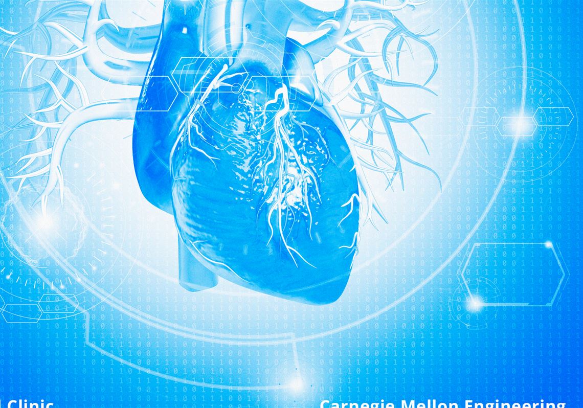
The article highlights a significant advancement in medical technology: the application of artificial intelligence to cardiac Magnetic Resonance Imaging (MRI). Cardiac MRI is recognized as the 'gold standard' for comprehensively assessing heart structure, function, and tissue health. However, its interpretation is an incredibly specialized and time-intensive task. Clinicians often dedicate at least 40 minutes to meticulously reviewing hundreds of images and manually annotating various aspects of each exam. This high demand for expert interpreters, coupled with a limited supply of such specialists, creates a bottleneck in patient care, underscoring the urgent need for innovative solutions that can support and augment human expertise.
To address the challenges in cardiac MRI interpretation, researchers from Carnegie Mellon University and the Cleveland Clinic's Cardiovascular Innovation Research Center have developed an innovative AI system named CMR-CLIP. This sophisticated system was rigorously trained using an extensive dataset comprising 13,000 patient studies and over a million individual images collected over a decade at the Cleveland Clinic. Unlike traditional AI models that often rely on manually labeled data, CMR-CLIP was specifically designed to learn by aligning MRI image sequences with existing natural language clinical summaries and radiology reports that physicians routinely complete for every patient exam. This approach leverages readily available clinical documentation to teach the AI how medical professionals describe and interpret scans in real-world practice.
A core challenge in developing AI for medical imaging is the scarcity of expert-annotated data, which is time-consuming and expensive to produce at scale. The CMR-CLIP team ingeniously circumvented this limitation by utilizing radiology reports already embedded in routine clinical workflows as its primary training data. This method allowed the AI to learn directly from the nuanced interpretations of human experts without requiring new, dedicated annotations. Consequently, CMR-CLIP demonstrated superior performance, interpreting complex heart scans up to 35% better than existing general-purpose AI models. Furthermore, the system exhibited a remarkable ability to identify conditions it had not been explicitly trained on, simply by matching images to descriptive prompts such as 'enlarged left ventricle,' showcasing its advanced understanding and generalization capabilities.
Beyond static image analysis, CMR-CLIP significantly advances cardiac AI by representing MRI exams as dynamic videos rather than individual frames. This innovative approach enables the system to monitor not only the static structure of the heart but also its movement and function over time, mirroring the comprehensive diagnostic process undertaken by human clinicians who interpret images from multiple angles and time stamps. According to Ding Zhao, associate professor in CMU's Department of Mechanical Engineering and co-principal investigator, this domain-specific model design allows it to 'unlock new levels of performance and clinical utility' compared to generic AI systems. The researchers further validated CMR-CLIP's robustness and generalizability by successfully testing it on entirely separate datasets collected in France and at Cleveland Clinic Florida, proving its potential for widespread adoption across different hospital systems globally.
The development of CMR-CLIP holds substantial promise for transforming clinical workflows and improving patient access to critical diagnostic technologies. Dr. David Chen of Cleveland Clinic, a co-principal investigator, emphasized that systems like CMR-CLIP have the potential to provide crucial support to clinicians through automated screening and interpretation assistance. This is particularly vital in settings where the availability of expert readers is limited, ensuring that more patients can benefit from powerful diagnostic technologies like cardiac MRI. By reducing the time and specialized expertise required for interpretation, CMR-CLIP can help accelerate diagnoses, potentially leading to earlier intervention and better health outcomes for individuals with heart conditions, thereby making advanced cardiac care more accessible and efficient.