Quick Buzz Feed

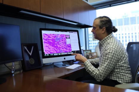
When a patient needs a second opinion, how does the biopsied tissue travel? The answer is surprisingly analog for 2026. Glass slides containing biological materials are often carried between offices so multiple pathologists can take a look. According to Lee Cooper, director of computational pathology at Northwestern University’s Feinberg School of Medicine, digital imaging methods are becoming more popular in the field of pathology. After a sample of blood or tissue is taken from a patient and placed on a glass slide, the slide is inserted into a scanner that produces a digital image that can be shared electronically. In addition to enabling frictionless travel, digitized samples can be reviewed by artificial intelligence and machine learning algorithms. Cooper, who runs a lab in which researchers develop such algorithms, says the algorithms will help pathologists make more accurate and detailed diagnoses, as well as help researchers interpret how diseases have progressed in past patients.
Researchers in the Cooper Lab are focused on developing and implementing artificial intelligence and machine learning algorithms to interpret digital pathology images. Their work involves writing these algorithms, digitizing biological specimens, and meticulously analyzing the resulting high-resolution images. This analysis is conducted on both ongoing research study samples and historical, previously banked samples, primarily to retrospectively understand disease progression in patients. Each researcher within the lab specializes in a unique dataset or a particular challenge faced by pathologists. For instance, Fattaneh Pourakpour, a Ph.D. student, develops AI algorithms for tissue segmentation, differentiating various brain regions like gray and white matter, and for cell classification within gray matter, especially using data from a brain bank focused on dementia and Alzheimer's disease. Her research also includes feature engineering to uncover new insights and identify potential biomarkers for neurodegenerative disorders. Similarly, Ramin Nateghi, a post-doctoral researcher, applies machine learning to detect prostate cancer, emphasizing the algorithms' ability to identify subtle cancerous changes that are often imperceptible to the human eye, thereby assisting doctors in making more informed and timely diagnostic decisions, especially in slow-progressing conditions.
The primary goal of digital pathology tools is to augment, rather than replace, the work of human pathologists. Lee Cooper highlights that completely replacing human expertise is not feasible, partly due to the existence of complex and rare 'edge cases' that current correlation-based AI models may struggle with due to a lack of causal understanding. While algorithms can process and consider a vast number of properties in a sample—far more than a human can—leading to more comprehensive predictions about a patient's future health, Cooper cautions against implicit trust in these models. The medical field, particularly pathology, generates an enormous volume of data; for example, Northwestern Medicine produces approximately 2 million glass slides annually. Compounding this is a significant shortage of pathologists in the United States, meaning there is an overwhelming amount of patient biopsy data requiring inspection and analysis. Researchers like Lawrence Chillrud, a Ph.D. student, are developing tools specifically designed to maximize the efficiency and accuracy of pathologists. These tools can perform preliminary screenings to categorize straightforward cases, flag complex ones for expedited specialized review, help distribute workload among pathologists, count specific cellular components, direct pathologists' attention to critical areas of a sample, and even review human reports for potential errors.
A critical aspect of AI development is the principle of 'garbage in, garbage out,' meaning that inherent flaws or biases in the training data will be reflected in the model's predictions. Lawrence Chillrud's research addresses this by developing 'uncertainty tools' that quantify the reliability and potential error rate of a model's prediction. These tools are vital because AI models can often be 'overconfident,' presenting an overly optimistic view of their accuracy. By flagging results with a high probability of error, these tools provide pathologists with crucial context, indicating when a model's predictions might need extra scrutiny or should be disregarded. Furthermore, uncertainty tools play a significant role in mitigating bias in artificial intelligence. If a model is trained predominantly on data from a homogenous demographic (e.g., white, affluent patients), its predictive accuracy may be compromised when applied to patients from different backgrounds. Uncertainty tools can alert pathologists when a prediction might be affected by gaps or biases in the model's knowledge base. Beyond bias, AI pathology tools also offer a solution to the shortage of specialized pathologists. They can empower general pathologists to handle cases like prostate cancer biopsies that typically require specialized training, making advanced diagnostic support more accessible, especially in underserved or rural areas.
Despite the undeniable potential and numerous benefits of digital pathology, its widespread integration into routine medical practice faces several significant obstacles. One major hurdle is the rigorous FDA clearance process required for digital pathology tools, which demands extensive validation with exceptionally high-quality data—data that can be challenging to acquire and standardize. Another limitation is the varying adoption rates of digitizing tools and technology across different medical systems; not all facilities possess the advanced infrastructure available at institutions like Northwestern. The sheer volume of data generated by digital pathology also presents a considerable challenge. For instance, the digitization of pathology slides at Northwestern alone can produce approximately 3 petabytes (3 million gigabytes) of data annually, necessitating robust storage, processing, and management solutions. Despite these challenges, Lee Cooper remains optimistic about the future of digital pathology. He observes a growing trend where not just tool developers, but also a broader community of scientists, are actively utilizing these technologies to measure intricate patterns in tissue and correlate them with critical clinical outcomes, such as the success of various treatments. This increasing adoption by the scientific community, and the demonstrated utility and ease of use of these tools, fuels his positive outlook on their eventual commonplace adoption.