A new experimental artificial intelligence (AI) system, called OCTCube-M, developed by researchers at Washington University School of Medicine in St. Louis, in collaboration with the University of Washington and Genentech, Inc., is designed to significantly improve and expedite the diagnosis of retinal diseases. This advanced technology analyzes detailed three-dimensional images of the eye's retina obtained through non-invasive optical coherence tomography (OCT) scans. The study, published in Nature Biomedical Engineering, demonstrates that OCTCube-M surpasses older 2D models in accurately identifying eight different retinal conditions, including age-related macular degeneration, the leading cause of blindness in individuals over 50. Beyond improved diagnosis, the AI system also shows greater precision in predicting the progression rate of severe conditions like geographic atrophy. This innovation promises to empower physicians with faster, more accurate diagnostic capabilities, refine treatment strategies, and accelerate the development of new therapies through more efficient clinical trials. Furthermore, the model exhibits the potential to detect broader systemic health risks, such as heart attack, stroke, and kidney failure, by analyzing subtle patterns in retinal blood vessels, thereby transforming routine eye exams into powerful tools for comprehensive health screening and early disease detection.
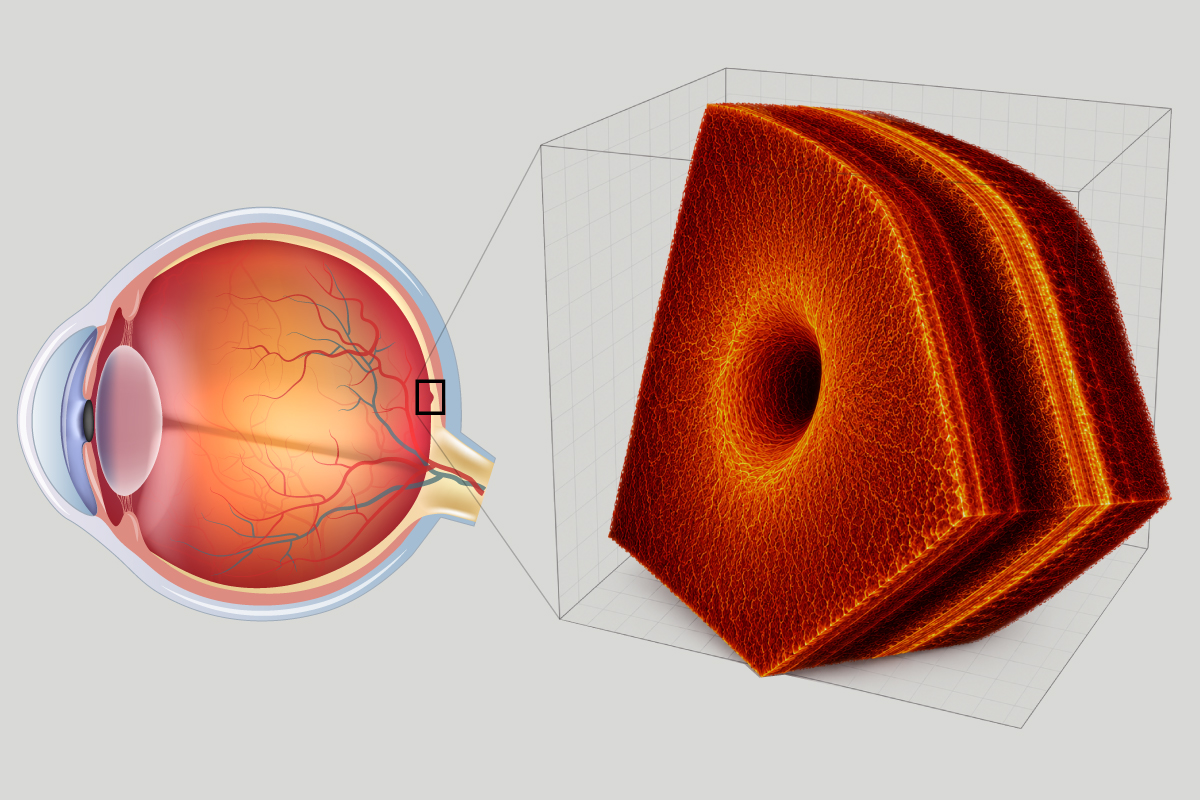
A diagnosis needle in a haystack of data
The advent of non-invasive optical coherence tomography (OCT) scans has revolutionized ophthalmology by offering physicians a highly detailed, three-dimensional view beneath the eye’s surface. These scans generate hundreds of cross-sectional images per patient, creating a vast and complex dataset that requires manual review by ophthalmologists. This laborious process is not only time-consuming but also susceptible to human error, posing a significant challenge in the timely and accurate diagnosis of various vision-threatening conditions such as glaucoma, macular degeneration, and diabetic retinopathy. Recognizing this challenge, researchers, including Aaron Lee and Cecilia S. Lee from WashU Medicine, Sheng Wang from the University of Washington, and Miao Zhang from Genentech, built upon previous work that demonstrated AI's capability in diagnosing eye diseases from 2D retinal images. Their latest endeavor focused on enhancing diagnostic precision by incorporating 3D tomography images, hypothesizing that a more complete, volumetric view of the retina would yield superior results, especially since many diseases manifest across three dimensions around the fovea, the critical area for sharp vision. To rigorously train this new system, named OCTCube-M, a massive dataset was compiled, consisting of over 26,000 3D OCT images, which collectively comprised 1.62 million individual retinal slices. Upon evaluation, OCTCube-M demonstrated a notable improvement in accuracy, identifying six out of eight retinal diseases four to six percentage points more accurately compared to its 2D predecessors. This translates to the potential detection of 43 to 60 additional cases per 1,000 individuals, showcasing its robust performance across diverse clinical sites, imaging modalities, and patient demographics. The model's ability to identify serious conditions affecting the back of the eye is particularly significant, as these are primary causes of vision loss and are frequently associated with broader systemic health issues like diabetes, hypertension, and cardiovascular disease. Further advancements involved integrating data from two additional eye imaging techniques—infrared retinal imaging and fundus autofluorescence imaging—into the OCTCube-M model. This multimodal approach enabled the AI to construct an even more comprehensive understanding of the eye's internal state. This enhanced version proved exceptionally adept at predicting the growth rate of geographic atrophy, a severe form of macular degeneration affecting approximately 5 million people globally, where effective treatment options are limited. The multimodal OCTCube-M outperformed existing state-of-the-art models that relied solely on fundus autofluorescence images by nearly 50%. This predictive capability is crucial for designing more efficient and targeted clinical trials for new therapies, potentially reducing costs, shortening development timelines, and accelerating the availability of effective treatments for patients. The researchers plan to continue expanding OCTCube-M's training with even larger and more diverse datasets, encompassing more patients, diseases, and imaging modalities, to further refine its capabilities and maximize its clinical utility.