Quick Buzz Feed

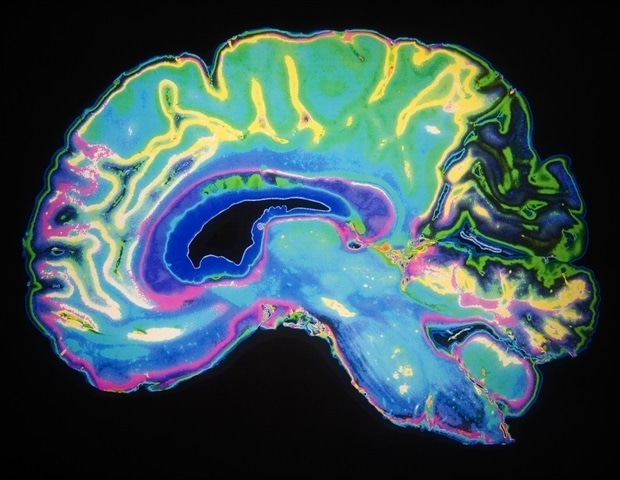
One of the uncomfortable truths about multiple sclerosis is that the part of the brain likely to reveal the most about the disease and how a patient will be impacted has been mostly invisible to clinicians.
For a long time, medical professionals have been intensely frustrated by the inability of standard Magnetic Resonance Imaging (MRI) to detect cortical lesions in multiple sclerosis (MS) patients. These gray matter lesions are crucial for understanding how the disease progresses and its impact on cognitive function. Traditional MRI techniques have only been effective in identifying white matter lesions, leaving a significant portion of the disease's ongoing damage unobserved in clinical settings. Despite decades of histopathological evidence from postmortem tissue confirming the presence and importance of these cortical lesions, clinicians lacked a practical method for their detection and monitoring in living patients. This new AI model marks a substantial advancement, offering a solution to this longstanding problem. Researchers emphasize that this collaborative effort represents a major success in applying artificial intelligence to medicine, finally unlocking access to vital diagnostic data from existing MRI scans that were previously hidden due to the limitations of conventional imaging, demonstrating the powerful capabilities of computational methods in medical diagnostics.
The research team successfully employed a sophisticated combination of multiple image-processing techniques, including a newly developed method called Multimodal Cortical Lesion Enhancement (MMCLE), to analyze existing MRI scans. This innovative approach was applied to a vast dataset of over 700 participants from the ORATORIO clinical trial, a large-scale, phase III FDA regulatory study focused on the MS drug Ocrelizumab. While individual MRI scans traditionally yielded visible evidence primarily of white matter lesions, the AI-powered processing of multiple different contrast images per patient revealed a remarkable number of previously undetectable cortical lesions. Specifically, the analysis identified an average of 15 to 20 cortical lesions per patient, accumulating to more than 11,000 across the entire study cohort. Dr. Michael G. Dwyer, the study's first and corresponding author, highlighted that generative AI's strength lies in its ability to discern minute differences between scans, which are imperceptible to the human eye. These subtle discrepancies signal abnormal tissue behavior, effectively bringing the hidden pathology of cortical lesions into view by synthesizing information across multiple MRI images that was otherwise missing.
The groundbreaking findings from this study are poised to profoundly influence future clinical trials and provide new insights into past trial data for multiple sclerosis. The ability to visualize and quantify what was previously considered 'invisible pathology' in the brain, particularly cortical lesions, is expected to significantly enhance understanding of disease progression and treatment efficacy. This success is a testament to the extensive collaboration between an international team of scientists and clinicians from both academia and industry. The University at Buffalo spearheaded the academic contributions, with key co-authors including Robert Zivadinov, Michael G. Dwyer, Niels P. Bergsland, Alexander Bartnik, and Dejan Jakimovski. Crucial industry partnership came from Genentech Inc., the pharmaceutical company behind Ocrelizumab, which also provided partial funding for the research. The interdisciplinary nature of this project, bringing together diverse expertise, was instrumental in achieving this significant advancement in MS diagnostics and research.